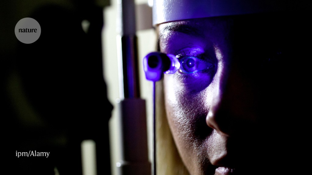
Eye disease and the risk of Parkinson’s can be detected by artificial intelligence
by admin

Evaluating Neural Tissues using a Large-Scale Model of Retinae Prediction and Textual Synthesis
Retinas are also an extension of the central nervous system, sharing similarities with the brain, which means that retinal images can be used to evaluate neural tissue. Some people don’t have the expertise to read the scans. This is where the computer-generated intelligence comes in.
Instead, the scientists used a method similar to the one used to train large-language models such as ChatGPT. The tool uses examples of human-generated text to learn how to predict the next word from the context of the preceding words. RETFound uses hundreds of photos of the retina to learn how to predict what parts of an image should look like.
Pearse Keane, an irish physician at Moorfields Eye Hospital Foundation Trust in London, co-authored a paper about the tool that learns what a retina looks like from millions of images. It is the cornerstone of the model, and this makes it a foundation model, which means that it can be adapted for many tasks.
The only part of the body which can be watched directly is the capillary network, the smallest of the blood vessels. “If you have some systemic cardiovascular disease, like hypertension, which is affecting potentially every blood vessel in your body, we can directly visualize [that] in retinal images,” Keane says.
Towards ethical and safe medical foundations for disease detection and diagnosis using real-time multidimensional images in the case of MRI and computed tomography
Xiaoxuan Liu, a clinical researcher who works in responsible innovation in artificial intelligence at the University of British Columbia, says that using unlabelled data to initially train the model blocks a major roadblock for researchers. The director of the center for artificial intelligence in medicine and the sciences agrees, according to a radiologist. “High-quality labels for medical data are extremely expensive, so label efficiency has become the coin of the realm,” he says.
RETFound techniques could be adapted to other types of medical scans, researchers are looking ahead. Langlotz is interested to see if the methods can generalize to more complex images like magnetic resonance images or computed tomography scans.
The authors have made the model publicly available, and hope that groups around the world will be able to adapt and train it to work for their own patient populations and medical settings. They could use data from their own country to fine-tune the way they use it.
“This is tremendously exciting,” Liu says. But using RETFound as the basis for other models to detect diseases comes with a risk, she adds. Limitations in the tool could cause models built from it to leak. “It is now up to the authors of RETFound to ensure its ethical and safe usage, including transparent communication of its limitations, so that it can be a true community asset.”
This work introduces a new SSL-based foundation model, RETFound, and evaluates its generalizability in adapting to diverse downstream tasks. RETFound can be adapted to a broad range of disease detection tasks and has been shown to improve performance for detecting cardiovascular and neurodegenerative diseases. It is a medical foundation model that has been developed and assessed, and shows considerable promise for leveraging such multidimensional data without constraints of enormous high-quality labels.
There is a big fall in performance when models are tested against new cohort’s different demographic profiles and also on the machines used for the evaluation phase This phenomenon is also seen in the evaluation of the diagnosis of ocular disease. 3b). The ischaemic stroke performance drops with the use of CFP and OCT. The drop in performance likely arises from the age and ethnicity profile of the internal and external validation cohort, as well as the imaging devices that are not the same. Compared to other models, RETFound achieves significantly higher performance in external evaluation in most tasks (Fig. 3b) as well as different ethnicities (Extended Data Figs. 9–11), showing good generalizability.
Researchers have developed a tool,Found, that uses hundreds of photos of the retina to learn how to predict what parts of an image should look like. “If you have some systemic cardiovascular disease…we can directly visualise [that] in retinal images,” Pearse Keane, a doctor at London’s Moorfields Eye Hospital, said. Found can be used to detect cardiovascular and neurodegenerative diseases.