DGC structure rationalizes muscular dystrophy-causing genes
Non-uniform refinement: adaptive regularization improves single particle cryo-EM reconstruction. Ann. Phys. 69 (2019)
Punjani, A., Zhang, H. & Fleet, D. J. Non-uniform refinement: adaptive regularization improves single-particle cryo-EM reconstruction. Nat. Methods 17 was published in 2020.
Chen, S. et al. It is possible to measure overfitting and validate resolution in 3D structure determination using a single particle electron microscope. Ultra microscopy was done at 24–35.
Muscular dissociation of a dystrophin-glycoprotein complex and its association with a C-terminal epitope
Durbeej, M. & Campbell, K. P. Muscular dystrophies involving the dystrophin-glycoprotein complex: an overview of current mouse models. The title was referred to as “cur.” Opin. Genet. There was a bill in 2002 that made it possible for people to have their licenses revoked.
Yoshida, M. et al. Dissociation of the complex of dystrophin and its associated proteins into several unique groups by n-octyl β-d-glucoside. The word eur comes from a Latin word. J. Biochem. 222, 1055–1061 (1994).
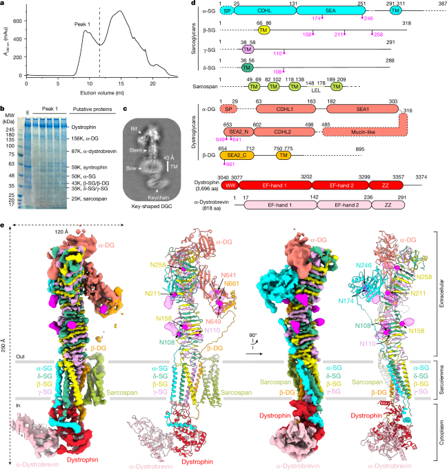
It’s F. et al. There is a dystroglycan binding epitope in the C-terminal region. The language of the European Union. J. Biochem. 268, 4590–4597 (2001).
Holt, K. H., Crosbie, R. H., Venzke, D. P. & Campbell, K. P. Biosynthesis of dystroglycan: processing of a precursor propeptide. FEBS Lett. 468 was published in 2000.
Duchenne Muscle Dystrophy Induced by a First Missense Mutation in the -sarcoglycan Gene
Mah, J. K. et al. There is a systematic review and meta-analysis on Duchenne muscular dystrophy. Neuromuscular Disord 24, 482–491 (2014).
Muthu, M., Richardson, K. A. & Sutherland-Smith, A. J. Implications for domain boundaries are given by the crystal structures of dystrophin and utrophin spectrin. PLoS ONE 7, e40066 (2012).
Ramaswamy, K.S. and others. Dystrophic mice and rats have muscles that are not able to transmit force. J. Physiol. 589, 1195–1208 (2011).
Marshall, J. L. et al. Dystrophin and integrin are important for sarcospan and a newly discovered muscle type in sarcospan-null mice. How could it be? Mol. Genet. 21, 4378–4373 was published in the year 2012
E. S. and Moreira have written about the causes of cancer in humans. A first missense mutation in the δ sarcoglycan gene associated with a severe phenotype and frequency of limb-girdle muscular dystrophy type 2 F (LGMD2F) in Brazilian sarcoglycanopathies. J. Med. Genet. 35, 951–953 (1998).
Piccolo, F. et al. Primary adhalinopathy—a common-cause of autosomal recessive muscular-dystrophy of variable severity. Nat. Genet. 10, 243–245 (1995).
M., saha, and others were involved in the project. There are correlations between PYROXD1 deficiency and muscular dystrophy in Saudi Arabia and Sudan. Physiol. Genomics 50, 929–939 (2018).
Piccolo, F. et al. A founder mutation in the γ-sarcoglycan gene of Gypsies possibly predating their migration out of India. Hum. Mol. There is a thing called Genet. 5 years ago.
Dai, Y. et al. Whole exome sequencing identified a novel DAG1 mutation in a patient with rare, mild and late age of onset muscular dystrophy-dystroglycanopathy. A cell. There is a difference between mol. Medical 23, 761–825.
Feng, J., Yan, J., Buzin, C. H., Towbin, J. A. & Sommer, S. S. Mutations in the dystrophin gene are associated with sporadic dilated cardiomyopathy. There is something called “mol.” Genet. Metab. 77 was published in 2002.
On the crystal structure of the endosialidase l-fucosidae of the marine bacterium Saccharophagus degradans 2-40
The marine bacterium Saccharophagus degradans 2-40 is a member of the polysaccharide degrading family. A man named J. Bacteriol. 193, 283–285 (2011).
Vickers, C. et al. -l-fucosidases fromGH29 have similar structural and mechanistic similarities to theendo-fucoidan hydrolases. J. Biol. Chem. 293, 18296–18308 (2018).
Both autoproteolytic and folding of the SEA domain of MUC1 mucin were found. Struct. Mol. Biol. 13, 71–76 (2006).
Miller, G., Wang, E. L., Nassar, K. L., Peter, A. K. & Crosbie, R. H. Structural and functional analysis of the sarcoglycan–sarcospan subcomplex. Exp. Cell. Res. 313 was published in 2007.
Stummeyer, K., Dickmanns, A., M., and Ficner,R. studied the crystal structure of the polysialic acid-degrading endosialidase. There was a systematic approach to the problem. Mol. Biol. The year 2004, 92–98.
Source: Native DGC structure rationalizes muscular dystrophy-causing mutations
The role of the tyrosine-phosphorylation and nonphosphorylated isoforms of the dystrophin-glycoprotein complex in the maintenance of scythe
R. M and Grady are involved in this case. There are tyrosine-phosphorylated and nonphosphorylated isoforms of -dystrobrevin. J. Cell Biol. 160, 741–752 (2003).
Grady, R. M. et al. There’s evidence for roles of the dystrophin–glycoprotein complex in the maintenance of the scythe. In 2000, Neuron 25, 279–289.
Ponting, C. P., Blake, D. J., Davies, K. E., Kendrick-Jones, J. & Winder, S. J. ZZ and TAZ: new putative zinc fingers in dystrophin and other proteins. Trends in medicine. There are articles in the Sci 21, 11–13 section of the journal.
Peter, A. K., Marshall, J. L. & Crosbie, R. H. Sarcospan reduces dystrophic pathology: stabilization of the utrophin–glycoprotein complex. J. Cell Biol. 183, 419–427 (2008).
Source: Native DGC structure rationalizes muscular dystrophy-causing mutations
Dystroglycan and dystrobrevin in Dag1-null mice. Its association with muscular dystrophy
Susa, K. J., Kruse, A. C. & Blacklow, S. C. Tetraspanins: structure, dynamics, and principles of partner-protein recognition. Trends Cell Biol. There is a publication in the Journal of Testing and Calibration (2023).
R. A. and Williamson wrote a book about it. Dystroglycan is essential for early embryonic development: disruption of Reichert’s membrane in Dag1-null mice. It’s a little dull. Mol. It was called Genet. A number 6 was produced in 1997.
Peter, A. K. The sarcoplasmic reticulum in the body is absent in the limb girdle muscular dystrophy 2F. Skelet. There is muscle 7 and 11.
Yoshida, M. et al. Evidence for an association of the sarcoglycan–sarcospan complex with dystrobrevin can be found in the biochemical evidence. That was very dull. Mol. The expression is also called Genet. 9, 1033–1040 (2000).
Source: Native DGC structure rationalizes muscular dystrophy-causing mutations
Crystal structure of the virulence factor P.69 pertactin from Bordetella pertussis (Astergillus aculeatus)
Yoder, M. D., Keen, N. T. & Jurnak, F. New domain motif: the structure of pectate lyase C, a secreted plant virulence factor. Science 260, 1503–1507 (1993).
Emsley, P., Charles, I. G., Fairweather, N. F. & Isaacs, N. W. Structure of Bordetella pertussis virulence factor P.69 pertactin. Nature 381 was published in 1996.
Petersen, T. N., Kauppinen, S. & Larsen, S. The crystal structure of rhamnogalacturonase A from Aspergillus aculeatus: a right-handed parallel β helix. Structure 5: 533.
Source: Native DGC structure rationalizes muscular dystrophy-causing mutations
Cryo-particle analysis in cryo-EM with a positive-unlabeled convolutional neural network: The case of strong sarcospan and as a musculoskeletal stabilizer
E.M., and his associates, collaborated on a project. High levels of sarcospan are well tolerated and act as a sarcolemmal stabilizer to address skeletal muscle and pulmonary dysfunction in DMD. Hum. Mol. Genet. 25, 5 395–5406.
Mitchell, R. D., Palade, P. and Fleischer present the purification of skeletal muscle structures. The J. cell Biol. 96 was published in 1983.
Kimanius, D., Dong, L., Sharov, G., Nakane, T. & Scheres, S. H. W. New tools for automated cryo-EM single-particle analysis in RELION-4.0. Biochem. J. 478, 4169–4185 (2021).
Bepler, T. et al. Particle picking in cryo-electron micrographs can be done with positive-unlabeled convolutional neural networks. Nat. Methods 16, 1153–1160 was published in 2019.
Rosenthal, P.B. and Henderson, R. Optimal determination of particle orientation, absolute hand, and contrast loss in single-particle electron cryomicroscopy. J. Mol. The book was 721–745.
Source: Native DGC structure rationalizes muscular dystrophy-causing mutations
Abramson and his paper Molecular Structure Prediction for AlphaFold 3 (2019): all-atom structure validation for macromolecular crystallography
J.Abramson and his paper. Accurate structure prediction of biomolecular interactions with AlphaFold 3. Nature https://doi.org/10.1038/s41586-024-07487-w (2024).
Chen, V. B. and others. MolProbity: all-atom structure validation for macromolecular crystallography. Acta Crystallogr. D. Crystallogr. 66, 12–21 (2010).
Scientists have developed an adaptive regularisation technique for single-Tesla cryo-electron microscopy that allows them to measure and validate resolution in the 3D structure determination using a single-particle electron microscope. They used the technique to demonstrate that it improves the resolution of 3D structure determination in cryo-EM reconstruction. The technique is used to measure dissociation of the complex of dystrophin and its associated proteins into several groups.