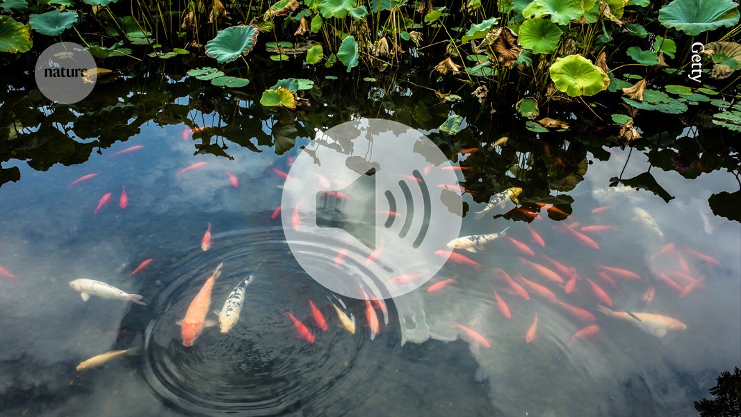
Scientists have an answer to how fish know where music comes from
by admin

Anomalous Krause corpuscles activate sexual behaviours in the penis and clitoris using microscopic nerve cells
150 years after they were discovered, researchers have identified how specific nerve-cell structures on the penis and clitoris are activated. These structures are similar to touch-activated corpuscles on people’s fingers and hands but there is little known about their role in sex or how they work. Working in mice, a team found that Krause corpuscles in both male and females were activated when exposed to low-frequency vibrations and caused sexual behaviours like erections. The researchers hope the work will help them understand the neurological basis for certain sexual problems.
Astronomers are unsure of the origin of a mysterious object and how gene editing could make rice plants more water efficient.
Behavioural Observation of the Pressure and Motion of a Fish Using a Particle Image Velocimetry Toolbox
To estimate binaural cues in our behavioural experiment, we analysed the pressure and particle motion at sound calibration grid points 3 cm apart, (({x}{0})) 1.5 cm to the left and (({x}{1})) 1.5 cm to the right of the centre grid point. To estimate sign and peak amplitude of level differences, we calculated P-ILD (pressure ILD) as ({\max }{t}({\rm{abs}}(p({x}{0},t)))\,-{\max }{t}({\rm{abs}}(p({x}{1},t)))) and M-ILD (particle motion ILD) as ({\max }{t}({\rm{abs}}({a}{x}({x}{0},t)))\,-{\max }{t}({\rm{abs}}({a}{x}({x}{1},t)))). The level differences between these two points were divided by a factor 50 to estimate the level difference across the left-to-right inner ear axis of the fish (about 0.6 mm). Comparing the single-speaker configuration with the trick configuration, these data show that the sign of M-ILD remains the same (+0.11 m s−2 versus +0.30 m s−2), but the sign of P-ILD is inverted (+4.4 Pa versus −4.6 Pa). The extended data fig. depicts the equation of P-ILD in a geometric fashion. 8d.
Don’t miss an episode. Subscribe to the Nature Podcast on
  Apple Podcasts


,


  Spotify


,


  YouTube Music


 or your favourite podcast app. An RSS feed for the Nature Podcast


 is available too.
To analyse the motion of the inner structures of the fish, we used Matlab 2019b and a particle image velocimetry toolbox PIVlab80, originally developed to characterize the motion of flowing particles for fluid mechanics. The particle displacement is assessed by a cross-correlating subregion with decreasing images. The contrast of the reflectance images was enhanced before the displacement analysis, and the results were curated in post-processing by removing outliers and interpolating detection gaps.
To reconstruct the relative phases and the amplitude of the object’s motion, we needed to measure the pixel under two different phases. As noise can influence this measurement, we used four phase steps here, ensuring proper phase reconstruction while keeping acquisition sessions reasonably short.
A new two-photon laser-scanning vibrometry experiment with untreated line-ablated fish (Extended Data Fig. 10b)
The principle of the laser- scanning vibrometric measurement is shown in fig. 3b and Extended Data Fig. 10b. The sample (Extended Data Fig. 10b(i)) was stimulated with an acoustic sinusoidal wave at frequency ({f}{{\rm{stim}}}), and imaged with a laser-scanning microscope with a line rate ({f}{{\rm{scan}}}) (Extended Data Fig. 10b(ii)
Fish were anaesthetized in 120 mg l−1 fish water-buffered MS-222. They were subsequently placed on a preformed agarose mould, which allowed the gill covers to move freely, and immobilized with 2% low-melting-point agarose (melting point 25 °C). The flow of aerated aquarium water was delivered through a glass capillary.
The microscope was built from a laser-scanning two-photon microscope. The source of illumination was a Ti:sapphire laser. The beam was transmitted through a 90:10 beam splitter before entering the microscope. The light back-scattered by the fish inner structures was descanned, reflected by the 90:10 beam splitter, and then focused by a lens (f = 50 mm) into a single-mode fibre (core diameter: 25 µm, numerical aperture: 0.1) acting as a confocal pinhole. The microscope was controlled by custom-written software (https://github.com/danionella/lsmaq).
The data in Figs. 1–4 and Extended Data Figs. 4–7 and 12a stem from 65 untreated fish (3,798 playbacks, 1,415 startles, about 37% startles). The number of fish that responded with at least one startle was indicated. The same experiment, also comprising 12 sound configurations, was repeated with 74 lateral line-ablated fish (Extended Data Figs. 7 and 9; 2013 playbacks, 910 startles, 45% startles). A third sound playback experiment was carried out in the dark in 43 untreated fish, testing a subset of 4 sound configurations (Extended Data Fig. 12b).
A 12-month-old male wild-type D. cerebrum was euthanized by ice shock and fixed with 4% paraformaldehyde in phosphate-buffered saline (PBS) at 4 °C overnight. The next day, the fish was washed for 15 min in PBS before being stained with 5% phosphomolybdic acid (Sigma Aldrich) solution in PBS at 4 °C overnight. After staining, the fish was washed in PBS for 15 min before embedding in 1% PBS-buffered agarose inside a cryo tube. The micro-CT scan was carried out at the ANATOMIX beamline at SOLEIL synchrotron by XPLORAYTION. The X-ray beam was white and the sample was placed there. An effective total size of 0.6485 m was achieved thanks to the 3,200 projections collected at about 10 optical magnification and a sensor size of 6.5 m. The registered data were binned to 1.2970 µm voxel size. The hearing apparatus’ key structures were manually segmented. To this end, the planes were hand-labeling using 3D Slicer76 and then interpolated using Biomedisa. The files were converted between different file types with the aid of the FIJI ImageJ. The segments were turned into mesh grids and loaded into Blender for cleaning and rendering.
Directional bias across trials and fish. For each stimulus or set of stimuli, startle trials were pooled across all fish, and the fraction of startles in one direction was calculated. The two-sided binomial test was used to calculate how likely a measured approach would have been if the response was not biased.
Pose tracking of D. cerebrum’s swimming behaviour was carried out with SLEAP75. 140 frames of random recordings of both male and female fish were hand-labelled, with a skeleton consisting of 7 equidistant points on the body segments and 2 additional points for each eye. A single animal model was used for training. The model parameters and the trained model are available at the G-Node repository (see Data availability).
Each fish was tested once and then again at a later time. In the first minutes of the recording, a 10 cm × 10 cm acrylic plate with centimetre markings was placed in the inner tank to match the sound calibration grid to the video frame. Three minutes after placing the fish into the inner tank, the water went back up.
The LAGeSo, the Berlin authority for animal experiments, approved all animal experiments that conformed to Berlin state, German federal and European Union animal welfare regulations. The aquarium that D. cerebrum was kept in had pH 7.3, 350 S cm1, and temperature 27 C. We used male and female fish for a long period of time.
Source: The mechanism for directional hearing in fish
Mapping the particle acceleration at a distance from a sound monopole using a discrete Fourier transform and a volumetric complex map
To compute the particle acceleration at a distance from a sound monopole with pressure we used this. Given a pressure waveform ({{{\bf{p}}}{n}}\,:={p}{0}{,p}{1},\cdots ,{p}{N-1}) with (N) samples ({p}{n}), spaced at (T=1/{sr}) with sample rate ({sr}), the particle acceleration ({{{\bf{a}}}{n}}\,:={a}{0}{,a}{1},\cdots ,{a}{N-1}) that would be observed at a distance (r={r}{0}) from a sound monopole was calculated by carrying out the discrete Fourier transform ({{{\bf{P}}}{l}}\,:={P}{0}{,P}{1},\cdots ,{P}{N-1})
This in turn set additional constraints on the various scanning parameters. We used (f_rmstim), which is the data presented in the picture.
The motion detection yielded x- and y-displacement maps at each of the four phases in the acoustic stimulation period. The first Fourier component was used to find the phase and amount of the displacement. The phase was finally corrected for the accumulating phase offset along the horizontal x direction due to the line scanning procedure (Extended Data Fig. 10c). The motion response of the various inner structures to the acoustic stimulation could be mapped with a consistent volumetric complex map by using this measurement in several planes. Maximum-amplitude projections across planes delivered the shown two-dimensional phase maps, one for motion along the speaker–speaker axis (x) and one for motion orthogonal to the speaker–speaker axis (y).
In the experiment, (r_0) was set to 3 cm so as to reproduce a monopole sound source at 3 cm, regardless of the speaker position. A peak particle acceleration of 7.59 m s2 was achieved. Other parameters are (c0.05),mathrm 1,500,rmm,rms-1. In terms of pressure, x acceleration and y acceleration (p, ax and ay), there were eight different target configurations, with ‘+’ indicating polarity of the template waveform and ‘−’ indicating opposite polarity: four monopole configurations (+,+,0), (−,−,0), (+,−,0) and (−,+,0); two pressure configurations (+,0,0) and (−,0,0); and two motion configurations (0,+,0) and (0,−,0). Despite a total of eight target configurations, there were 12 sound configurations as the four monopole configurations can be realized in two ways, either with a single speaker or with three speakers (trick configuration; see the next section).
wavelength, speed of sound, and wave number are also included.
In a medium of density (\rho ), the radial particle velocity decays quadratically with distance in the near field (({kr}\ll 1), limit dependent on frequency):
Particle acceleration decays with distance for nearby sounds, and the limit is independent of Frequency.
In the second method, the particle acceleration was measured along all three axes with an instrument bought from the National Instruments. 1d Like the hydrophone, the acceleration sensor was moved across all 5 × 5 grid positions during repeated playback of the same sound, giving measurements for x, y and z acceleration.
Hence, in all experiments, x and y acceleration were measured through the indirect method, on the basis of spatial pressure gradients. The vertical z acceleration in our setup is measured by the particle acceleration sensor.
Whereas hydrophones are manufactured and calibrated for underwater use, the particle acceleration sensor is not made to measure particle acceleration underwater and is meant to be glued onto the vibrating object. Owing to an acoustic impedance mismatch between metal and water, we expected the PCB sensor to underestimate particle acceleration.
We compared x and y acceleration waveforms for both measurement methods and found that the acceleration waveforms acquired through the direct method match the waveforms acquired through the indirect method after multiplication by a factor of about 2.4. The validity of the approximation in the indirect method is confirmed by a close match.
Source: The mechanism for directional hearing in fish
Controllability of Speakers in QCD with Angular Momentum and Gravitational Cascades (Preliminary Version)
To increase robustness of the solutions, they had to be forced to become close to the target signal to avoid speakers cancelling themselves. The solution of the system of equations was done with a least square solver. The bound ({B}{i,l}) was computed as a rescaling of the absolute Fourier components of the target pressure waveform ({P}{l})
Past s_i mathopsum.
in which (\gamma ) is fixed and scales pressure to voltage and ({\alpha }_{i}) is a rescaling parameter set independently for each speaker to give additional control over active speakers. In supplementary table 2 we list values for different sound configurations.
Researchers have studied the pressure and particle motion of lateral line-ablated fish using a particle image velocimetry toolbox. They analysed the motion of inner structures of the fish using Matlab 2019b and particle image velocimetry toolbox PIVlab80, originally developed to characterise the motion of flowing particles in fluid mechanics. The experiment was conducted in the dark in 43 untreated fish.