Rat cells repair mouse brains
by admin

Mice-colon and brain ‘organoids’ shed light on cancer and other diseases: Sergiu Pașca’s study of the Alzheimer’s disease
Organoids — particularly those made from human stem cells — sometimes reveal things that animal models can’t, says Sergiu Pașca, a neuroscientist at Stanford University in California and a co-author of one of the studies1. Pașca’s group studies Timothy syndrome: a genetic disorder involving autism, neurological problems and heart conditions that affects only a few dozen people in the world. Timothy syndrome is caused by a single mutation in a gene called CACNA1C, which encodes a channel through which calcium ions enter cells including neurons.
There aren’t good animal models for Timothy syndrome due to the underlying disease not always causing the same symptoms in rodents. He says it became clear to them that they’d need a way to test in their own body.
The brain organoids were used by the researchers to recreate the disorder. Stem cells from 3 people with a genetic condition that results in Timothy syndrome were cultured for about 250 days and eventually turned into brain organoids containing all the types of brain cells found in the cerebral cortex. The team injected the structures into the brains of rats to create a more realistic environment for the organoids. This made a system in which the researchers could test potential treatments for the disorder.
Human neurons have four different forms of this calcium channel, but only one of them is defective in Timothy syndrome. The researchers suggest that getting rid of the channel would allow it to be replaced by healthy channels.
Towards a chimaera by combining rat and mouse neuronal cells in a mouse colon cancer model using laser-activated gene loci
He says his group will need to prove the therapy is safe before they can try it on people in clinical trials. People would have to receive frequent injections for three months if the treatment is effective according to the researchers. Paca claims that the biological effects of the treatment would be reversed and any side effects would be short-lived.
To make a model of colon cancer, they engineered the cells to contain light-sensitive proteins attached to cancer-causing genes. They were able to use a blue laser to switch on the genes and cause the tumours to grow at certain locations in the organoid.
When the researchers injected the cancerous cells into mice, the tumours looked similar to those seen in human colorectal cancer. The organoids accumulated fewer tumours when the researchers restricted calories in their medium, which also happens in people with colorectal cancer.
Wu says that his laboratory now plans to use the technology developed for these studies to make chimaeras by transplanting cells from wild rodent species into lab mice. It is difficult to study wild rodents because they are hard to keep and breed in captivity. Stem cells can be injected into mouse blastocysts to study how other species’ brains develop and function.
How neurons connect with one another, and fire, makes integrating cells from two species complicated, says Kristin Baldwin, a neuroscientist at Columbia University in New York City. She says that neurosciences are not just Legos.
In a paper published by one of the teams on 25 April in Cell1, Baldwin, molecular biologist Jun Wu at the University of Texas Southwestern Medical Center in Dallas and their colleagues attempted to test this by mixing rat and mouse neuronal cells very early in the mice’s development.
First, they engineered the genes in a group of mice in a way that destroyed some neurons in the animals’ olfactory systems. This disrupted the circuits linking olfactory neurons in the nose with higher brain regions, leaving the mice unable to use their sense of smell to find mini-cookies that the researchers had buried in various places throughout the animals’ cages.
In a Cell paper published by the second team, also on 25 April2, Wu and his colleagues developed a more aggressive strategy for getting rat cells into a mouse’s brain. A group of mouse blastocysts were wiped out thanks to the use of a genetic-editing tool that cuts genes in multiple places. This is a gene that controls the development of the forebrain in an animal.
“There’s lots of fascinating biology to be learnt from this [rat–mouse] chimaera,” says Jian Feng, a physiologist at the University of Buffalo in New York. He’s not surprised that the rat cells followed the pace of the mouse’s developmental ‘clock’. In 2020, his group published a paper about a mouse embryo that it had engineered to contain up to 4% human cells5. The embryo began developing human red blood cells 17 days into gestation — much earlier than these cells develop in human embryos — suggesting that human cells, too, could follow the molecular directions of their host.
There is a mismatch in the development rates of the species. However, the teams found that the mouse brains developed at the same rate as they would normally, rather than at the slower pace at which a rat usually develops.
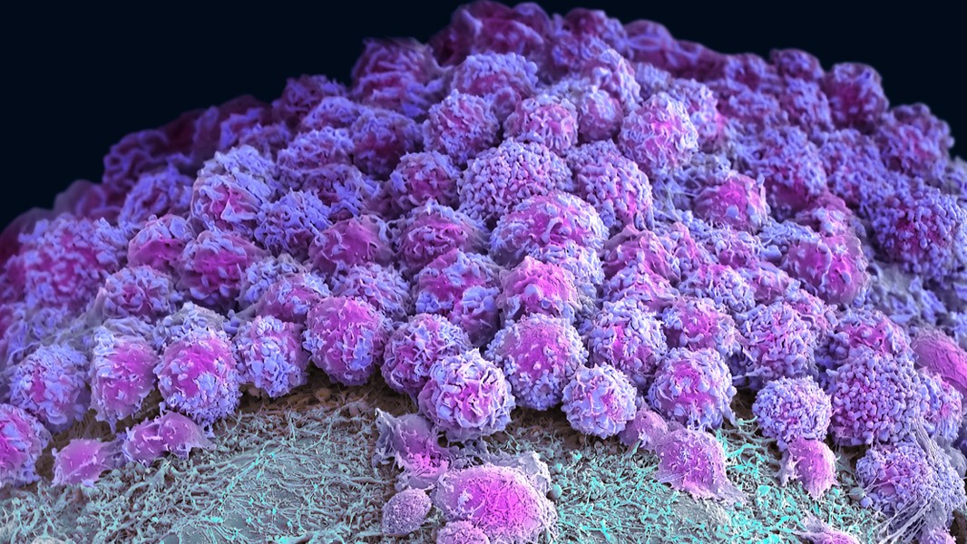
Researchers have created brain organoids from human stem cells cultured for 250 days, containing all types of brain cells found in the cerebral cortex. The organoids were injected into mice and revealed tumours similar to human colorectal cancer when the cancerous cells were injected into them. Researchers said that they plan to use the technology developed for these studies to make chimaeras.