The HIV capsid is similar to FG-nucleoporins
by admin

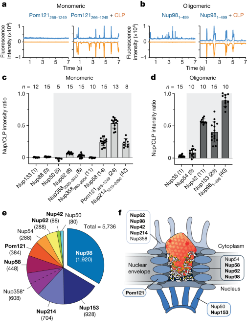
Nup 98 condensate-CA mixed experiments with HIV-1 fluorescent dyes using a Zeiss laser scanning confocal microscope
Lau et al.17 described FFS in detail. The cells-freeexpressed GFP-Nup and AF488 peptides were mixed with the HIV-1 CA assembly. Tris, pH 8, 150 mM Something like NaCl. Fluorescence traces were recorded for 15 s per trace in 1 ms bins using a scanning stage operated at 1 μm s−1. Normally, the measurements were repeated ten times per sample.
Owing to the length of Nup358, AlphaFold2 predictions were performed as three FG-containing sections (982–2004, 2005–3043 and 3058–3224), with their per-residue RSA-based disorder propensity calculated locally. Binary designations of order and disorder were assigned using a threshold optimized on Critical Assessment of Protein Intrinsic Disorder (CAID) data (0.581)57.
Experimental details for Fig. 3a–f and Extended Data Figs. 6 and 7: Nup98 condensates were immediately mixed with the following proteins: mCherry and IBB-mCherry (200 µM), importin-β:IBB-mCherry and importin-β + mCherry (both 100 µM), CA-mCherry and CA N57A-mCherry (45 µM), CAhexamer-mCherry and CA N57Ahexamer-mCherry (65 µM). The samples were imaged after around 5 min on the coverslip.
The image was performed using a 63 oil immersed objective and a Zeiss LSM 880 inverted laser scanning confocal microscope. There was a 180 5 m cover slide that was used to transfer the Nup98 reaction mixes into the Ibidi. Z-stacks were taken around the centre of the Nup98 condensates with the highest diameter. Images were processed using various programs.
Nup98 condensate-CA experiments were performed with mCherry-labelled CA (CA-mCherry and CAhexamer-mCherry), as it has previously been reported that fluorescent dyes such as Alexa568 can non-specifically interact with Nup condensates59.
Experimental details for Extended Data Fig. 8c–j: CA-mCherry and CAhexamer-mCherry (final 25 µM) were incubated with importin-α:importin-β, TNPO1 and TNPO3 (all final 2.5 µM), respectively, for 10 min, followed by mixing with Nup98 condensates and imaging after around 5 min incubation on the coverslip.
The largest diameter Z-slices were used to get the averaged radial intensity profiles. Profiles were removed. The edge of a condensate was defined from the 488 channel as the first intensity value greater than 5. The fluorescent bulb’s point spread function was taken into account when subtracting 200 NM from the value. In the radial averaged intensity graphs, this point was defined as point 0 m.
Unlabelled CLPs were prepared as described above. A two-step centrifugation protocol was performed as for confocal microscopy. Immediately before plunging to minimize sample aggregation, equal volumes of the suspension were mixed with shock-diluted condensates.
The mixture of freshly mixed Nup98 was applied to the Quantifoil R2/2 copper grid. The grid was exposed to a small amount of liquid ethane in the front by blotting it at 15 C and then using the Lecia EM GP device. The grids were cooled down in liquid nitrogen.
An electron microscope was used to image the grids. Cryo-ET data were collected with single-axis tilt on a Falcon III direct electron detector (Thermo Fisher Scientific) in linear mode at a magnification of ×28,000 with a pixel size of 5.23 Å. Tilt series were collected using the dose-symmetric scheme60 from −60 to 60° at 3° intervals using Tomography software (Thermo Fisher Scientific) with the defocus value set to −10 µm. Total dose for each tilt series ranged from 60–70 e/Å2. Before tomograms can be reconstructed, images of tilt series are binned. IMOD package61 was used to create three-dimensional reconstructions. The stack of tilted images were aligned using fidelity tracking.
There are pictures and graphs of confocal microscopy in extended data figs. The data is representative from at least three independent experiments. Confocal microscopy images in Fig. 4a and Extended Data Fig. 9 are representative of three independent experiments with similar results. There are graphs and images of frozen tissue. There are two independent experiments that have the same results shown in fig. 9.
Cell-free expression of GFP fusion proteins from E. coli rosetta2 cells using pCellFree03Nup plasmids
Cell-free expression of GFP fusion proteins was performed as described by Lau et al.17. RnaseOUT was added to Briefly Leishmania tarentolae extract. pCellFree_03-Nup plasmids (150 ng μl−1) were then added to the expression mix, followed by incubation at 27 °C for 3 h. The purity and degradation of the expressed Nup were assessed by a gel stain using sodium dodecyl sulfate and polyacrylamide.
Importin-α, TNPO1 and TNPO3 were expressed as GST-fusion proteins from E. coli Rosetta2 cells, as described above for importin-β and IBB-mCherry. Cell pellets were resuspended in 25 mM. Tris pH 7.5, 500 mM NaCl, 1 mM DTT, 10% glycerol and 1× cOmplete EDTA-free Protease Inhibitor (Roche) and lysed by sonication. Cell lysates were subjected to centrifugation (18,000g, 60 min, 4 °C). Clarified lysates were bound to GSH-resin and washed with 25 mM Tris pH 7.5, 150 mM NaCl, 1 mM DTT, 10% glycerol. PreScission was used to achieve oncolumn cleavage of thetag. The Cleaved proteins were subjected to SEC over the Superex 200 and equilibrated at 50 mM. Tris 7.5 pH and 200 mM. NaCl, 1 mM DTT, 10% glycerol. The frozenuted proteins were stored at 80 C.
To assemble the final cross-linked hexameric CAhexamer-mCherry, CAhexamer and CAhexamer-mCherry were mixed in a 5:1 ratio and assembled by means of the following dialysis steps: twice against 50 mM There is a tris pH 8, 1,000 mM. NaCl, 2 mM DTT; twice against 50 mM Tris pH 8, 1,000 mM NaCl; and twice against 50 mM Tris pH 8, 40 mM NaCl. The assembled CAhexamer-mCherry was finally purified with gel filtration (S200), and fractions containing hexameric CAhexamer-mCherry carrying one mCherry were pooled and snap frozen to be stored at −80 °C.
The untagged CAwt was over produced in E. coli cells. Cells were resuspended in a buffer of 50 mM. Tris/HCl pH 8.0, 50 mM NaCl, 4 mM TCEP, 2 mM PMSF and Turbo Nuclease). Cells were lysed and cleared using a 20,000gcentrifugation, then precipitated with a 25% saturation for 30 min. Precipitated protein was collected for 20 min by centrifugation. After the supernatant was thrown, the pellet was resuspended in lysis buffer and dialysed. The pH 8.0 and 1 mM are tris. There is a program called TCEP. CA was further purified by injecting on a HiTrapQ column and eluting it with 20 mM Tris/HCl pH 8.0, 100 mM 1 mM is NaCl. TCEP. The column was regenerated with 50 mM The tris/hcl has a pH of 8.0 and 2.0 M NaCl. TCEP to remove nucleic acid contaminations. The eluted CA was buffer exchanged by SEC (Superdex 200 10/300 increase) into 25 mM Tris/HCl pH 8.0. A group of Fractions were pooled and concentrated.
The HIV-1 CA proteins were expressed in Escherichia coli C41 or the satellite cells, N77V/A204C. HIV-1 CA K158C was mixed in with an unlabelled HIV-1 CA A 204C to create a 1:200 ratio. CA lattice assembly was carried out as described by Lau et al. 17 with the modification of a 15 min incubation at 37 °C and no overnight incubation. CAhexamer, CAhexamer-mCherry and CA-mCherry were purified based on previously described protocols27. Briefly, CAhexamer, CAhexamer-mCherry (pOPT) and CA-mCherry (pET21a) were expressed in E. coli C41 cells. While cells were growing at 37 C, both the inducer and cell growth were continued overnight at 18 C. Cells were collected (4,000g, 10 min, 4 °C), and the cell pellets were resuspended in lysis buffer (purification buffer 50 mM Tris pH 8, 50 mM NaCl, 2 mM dithiothreitol (DTT); +cOmplete EDTA-free Protease Inhibitor (Roche)) and lysed by sonication. The lysate was cleared by centrifugation (16,000g, 60 min, 4 °C), and an ammonium sulfate (20% w/v) precipitation was carried out on the soluble fraction. After stirring for 30 min at 4 °C, the precipitated material was pelleted (16,000g, 20 min, 4 °C). CAhexamer was further purified by resuspending the pellet in 100 mM citric acid pH 4.5, 2 mM DTT and dialysed against the same buffer three times. The precipitated protein was pelleted (16,000g, 20 min, 4 °C), and the soluble fraction was collected for further purification. CAhexamer-mCherry and CA-mCherry could not be purified with citric acid precipitation owing to a possible loss of mCherry fluorescence. The ammonium-sulfate-precipitated pellets were resuspended in 8 ml purification buffer per litre culture, run over a 20 ml Hi-TRAP Q column in purification buffer and eluted in a 1–100% gradient with buffer B (50 mM It has pH 8 and 500 mM. It’s a substance called NaCl. Fractions containing the same genes were pooled.
HeLa-K cells were obtained from the European Cell Culture Collection (RRID:CVCL_1922), authenticated by the manufacturer and tested negative for mycoplasma. Fetal calf and antibiotics were supplemented with DMEM, high-glucose, on 8-well -slides to 70%. The cells were treated with 30 g of digitonin in the transport buffer. 40 nM of anti-Nup133nanobodies47 and EGFP was used to put the cells in incubators for 30 min. Adding 3 M of a fragment with 1–180 and charged with GTP was indicated. The samples were then directly scanned with a Leica SP8 confocal laser-scanning microscope (equipped with a ×63 oil objective and HyD GaAsP detectors), using the 488 and 638 nm laser lines sequentially for excitation. Such live scan directly reads the distributions of the analysed probes between bulk buffer and NPCs—although local concentration at NPCs is underestimated because of the diffraction-limited resolution.
He14-bdSUMO-tagged CAP1A/N-21C/A22C was purified and concentrated to 15 thousandths of a liter. Assembly was performed in 50 mM There is a Tris/HCl with pH 8.0, 1 M NaCl and 0.1 mM Intellectual Property on hand. The capsid spheres were isolated from CA bycentrifugation at 280,000g. The pellet was washed and resuspended in assembly buffer. The spheres were further isolated by SEC on a Superose 6 10/300 column equilibrated in 50 mM. Tris/HCl pH 8.0, 500 mM NaCl and 0.5 mM IP6. Assembly could be performed with either 2 mM mCherry or with CA–sinGFP4a or CA–EGFP fusions, which is at one sixth of the non-fused CA.
The buffer contained 50 mM. Tris/HCl pH 8.0, 1 M NaCl, 100 µM IP6 (myo-inositol hexaphosphate) for 24 h at a protein concentration of 10–15 mg ml−1 at room temperature. The sample was then dialysed against 50 mM Tris/HCl pH 8.0. In 20 mM, the ethamers were isolated using a Superdex 200 increase column. Tris/HCl pH 8.0, 150 mM NaCl, 100 µM IP6. The samples were stored at an optimum temperature of 80 C for later use. There was validation of the purity of theprotein by an instrument.
Disulfide-stabilized CA hexamers were prepared as described in ref. 49. An amino-terminal His14 tag for Ni(II) chelate affinity purification and a Brachypodium distachyon (bd)SUMO tag65 were added for solubility to the CAP1A/A14C/E45C/W184A/M185A mutant. There was an amplification of expression in the cells. Liquid cultures were used to make a new culture. IPTG at 18 °C for 16 h. Cells were collected by centrifugation, resuspended in lysis buffer (50 mM Tris/HCl pH 8.0, 500 mM 20 mM imidazole, 1 mM. A high-pressure cell homogenizer was used to process TCEP, 2 mM PMSF. The lysate was cleared by centrifugation at 8,500g for 30 min. The conjugate was put to use with Ni Sepharose 6 Fast Flow beads for 1 h. The beads were subsequently washed with wash buffer (50 mM Tris/HCl pH 8.0, 40 mM imidazole, 300 mM NaCl, 1 mM TCEP). Proteins were eluted from the beads by tag cleavage with 2–5 µg ml−1 of bdSenP1 in cleavage buffer (50 mM Tris/HCl pH 8.0, 150 mM NaCl, 0.5 mM TCEP) for 3 h at 4 °C.
Supplementary Table 1 lists plasmids used for recombinant expression. In the references, expression and purification were detailed. 21,25,26,50,64).
Phase Separation of Finite-Gauge Growth Factors (FG) Particles from a Whole Nuclear Pore via a High-Process Fluorescence Transistor
Partition coefficients were used to divide the signal in independent FG particles by the reference areas in outside regions. The plots are for FG particles with 4-7 m diameter. Images were analysed in FIJI 2.9.0 and the exported data were further processed in GraphPad Prism 9.5.1.
The GLFG stock was dissolved in a mixture of 2 M guanidinium and 4 M guanidinium. Assay buffer for human Nup98-FGs and yeast Nup116-FGs was 50 mM The tris/hcl pH is 7.5. 1 mM is known as NaCl.
Minor changes were made to the assays. In brief, phase separation was initiated by rapidly diluting a 1 mM FG domain stock with 25 volumes (for GLFG) or 50 volumes (for the others) of assay buffer (50 mM pH 7.5 is the threshold for Tris/hcl. NaCl, 1 mM IP6), followed by another fourfold dilution in buffer containing the indicated fluorescent probes. FG particles and IbidI were allowed to be deposited on the slides for a short time before they were scanned.
Source: HIV-1 capsids enter the FG phase of nuclear pores like a transport receptor
Measurement of the dissociation of streptavidin conjugates in oocytes of 9-week-old CD1 mice using an Octet RED96 instrument
Mouse oocytes were obtained from ovaries of 9-week-old CD1 mice which were maintained in a specific pathogen-free environment according to the Federation of European Laboratory Animal Science Association guidelines and recommendations, as previously described53. The oocytes were kept in prophase in a homemade M2 medium and under the paraffin oil at 37 C. The capsid spheres were microinjected into the nucleus of oocytes. After the injection,ocytes were imaged in about 25 minutes.
A high-precision streptavidin biosensor tip was pre-incubated for 10 minutes. The tris/hcl was about 300 mM. NaCl, 0.1% (w/v) bovine serum albumin, 0.02% (v/v) Tween-20). Next, the conjugates prepared in a BLI buffer solution had to be conjugated to a thickness of 1 nm. Association was recorded for 180–300 s, followed by a 200–300 s dissociation step in wells containing BLI buffer. All binding sensorgrams were recorded on a forteBIO OctetRED96 instrument. A reference sensor was included and used to subtract background noise. Data were normalized to the baseline step and aligned to the dissociation start point using the Octet data analysis software. Data were plotted using PRISM.
An experimental method was developed using hexameric hexameric CAmer-mCherry and hexameric CASenmer-mCherry. The CAmer-mCherry was first incubated with importin-:importin-, TNPO1, TNPO3 (all final 2.5M). NaCl, 2 mM DTT, twice against 50 mM Tris pH 8, 1,000 mM NaCl, and twice against 50 mM Tris pH 8, 40 mM NaCl, and twice against 50 mM Tris pH 8, 40 mM NaCl, to remove nucleic acid contaminations.